Please click this link for pre-test.請點選此連結進行前測。
Chief complaint
Acute-onset of bloody stool over the course of one day.
Present illness
A 21-year-old male was presented to the emergency department with an acute-onset of bloody stool over the course of one day (Figure 1). The patient also reported simultaneous exertional dyspnea and dizziness. He recalled two episodes of painless bloody stool in the past 3 weeks, and colonoscopy had been performed twice without any specific bleeding being found. He denied any medical history related to his condition or any recent pharmacological use.
Figure 1. Bloody stool
 Past history
None.
Personal History
Smoking: Nil
Alcohol: Nil
Betel nut: Nil
Drug history: Nil.
Travel history
None.
Allergy history
Drug : Nil
Food : Nil
Blood transfusion : Nil
Family history
Significant disease history in the family: Nil.
Contact History
None.
Physical Examination
Upon physical examination at admission his temperature was 35.9℃, heart rate 107 beats/minute, respiratory rate 18 breaths/minute and blood pressure 90/51 mmHg. An abdominal examination revealed a hyperactive bowel sound during auscultation and a soft sensation while performing palpation without any muscle guarding or rebounding pain. The remainder of the physical examination was unremarkable with the exception of pale conjunctiva pallor.
Laboratory Data
A laboratory examination revealed a total leukocyte count of 17,560 cells per μL, Hemoglobin concentration of 7.3 g/dL, and platelet count of 429,000 per μL. The patient’s serum creatinine was 101.7 µmol/L, Internal Normalized Ratio (INR) 1.09, and activated Partial Thromboplastin Time (aPTT) 20.5 seconds on initial presentation. A chest X-ray examination was normal, while an electrocardiogram reported sinus tachycardia.
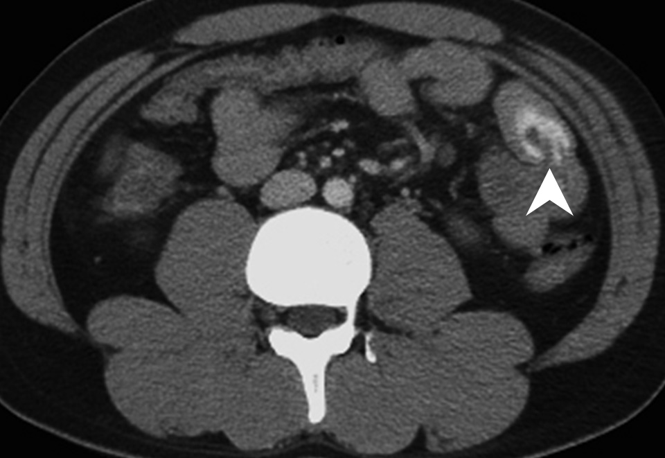
Computed tomography of the abdomen with biphasic contrast enhancement exhibited active contrast extravasations and segmental wall thickening in the jejunum (Figure 2).
Figure 2. Computed tomography of the abdomen
 2022/9/7 17:17:00
 873
873


