Please click this link for pre-test.請點選此連結進行前測。
Chief complaint
Shortness of breath for 2 weeks.
Present illness
Mrs. Yuan is a 65-year-old female patient denied systemic disease before. Her ADL was fair before. This time, she suffered from shortness of breath for 2 weeks with progression in recent 4 days. There was no fever, cough, chest pain, abdominal pain, limbs swelling, nor headache. The TOCC history was that she had been to Kaohsiung mountain area but denied insect bite. She called at CV OPD initially then was referred to Emergency Room where shallow respiration and rales breathing sound were noted. The CXR showed bilateral increased pulmonary infiltration. Bedside cardiac echo showed fair LV wall motion but moderate MR. Chest CTA did not reveal pulmonary embolism but right femoral vein DVT. Lab data: WBC 5590/μL, Hb 7.3g/dL, MCV 101.3 fL, platelet 146k/μL, albumin 2.4mg/dL. Under the impression of normocytic anemia, acute pulmonary edema and DVT, she was admitted.
Past history
Denied
Personal History
Smoking: Nil
Alcohol: Nil
Betel nut: Nil
Drug history: Nil
Travel history
Kaoshiung
Allergy history
Drug : Nil
Food : Nil
Blood transfusion : Nil
Family history
Significant disease history in the family: Nil
Contact History
No
Physical Examination
On exam, pale conjunctiva was noted. Right lower leg non-pitting edema. Otherwise, no specific findings.
Laboratory Data
Blood test showed :WBC:5590 /μL, Hb: 7.3g/dL, PLT: 146 x10^3/μL, Neut/Lym: 62%/25%,Metamyelocyte: 1%, Myelocyte: 1%, aty. Lym: 1%, albumin 2.4 g/dL, total protein 13.0 g/dL.
Images
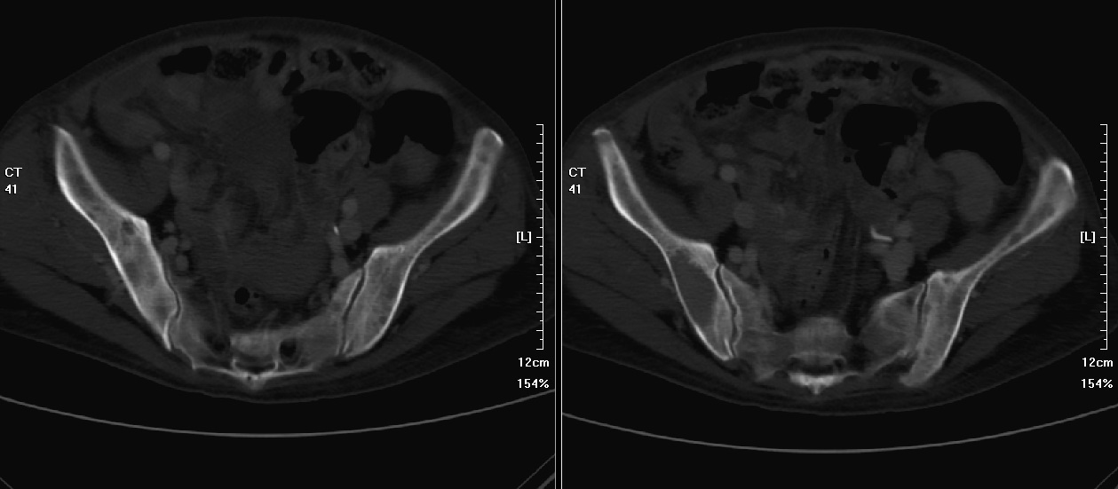
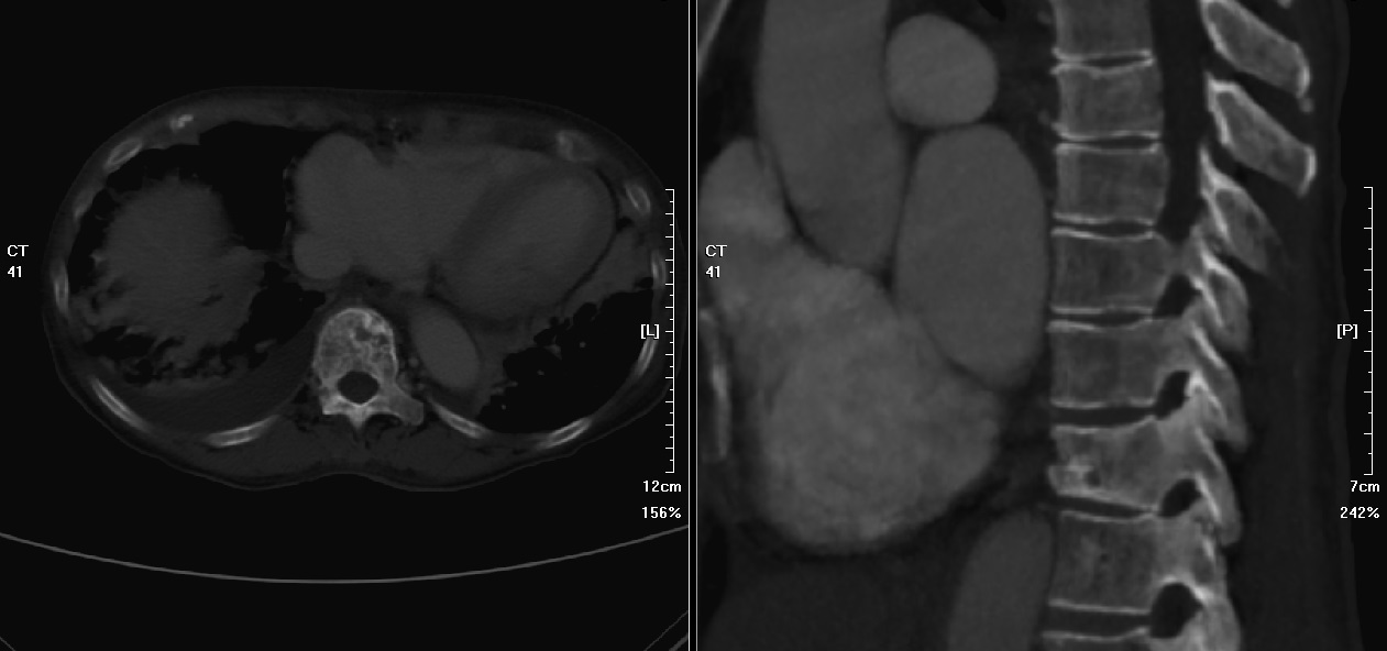
Contrast enhanced CT scan of chest, abdomen with CTA of pulmonary artery and with indirect CT venogram, showed multiple osteolytic bone metastases, to bony pelvis(Picture 1) and T11 body(Picture 2).
Metastatic bone lesions in pelvic bone(Picture 1)
 Metastatic bone lesion in T11 vertebral body(Picuture2)
 2022/9/7 17:23:42
 927
927


